
Chest X-Ray Outlier Detection, Classification Using Liquid Neural Networks with Dimension Reduction and Advanced Edge Detection
Article Sidebar
Main Article Content
Abstract
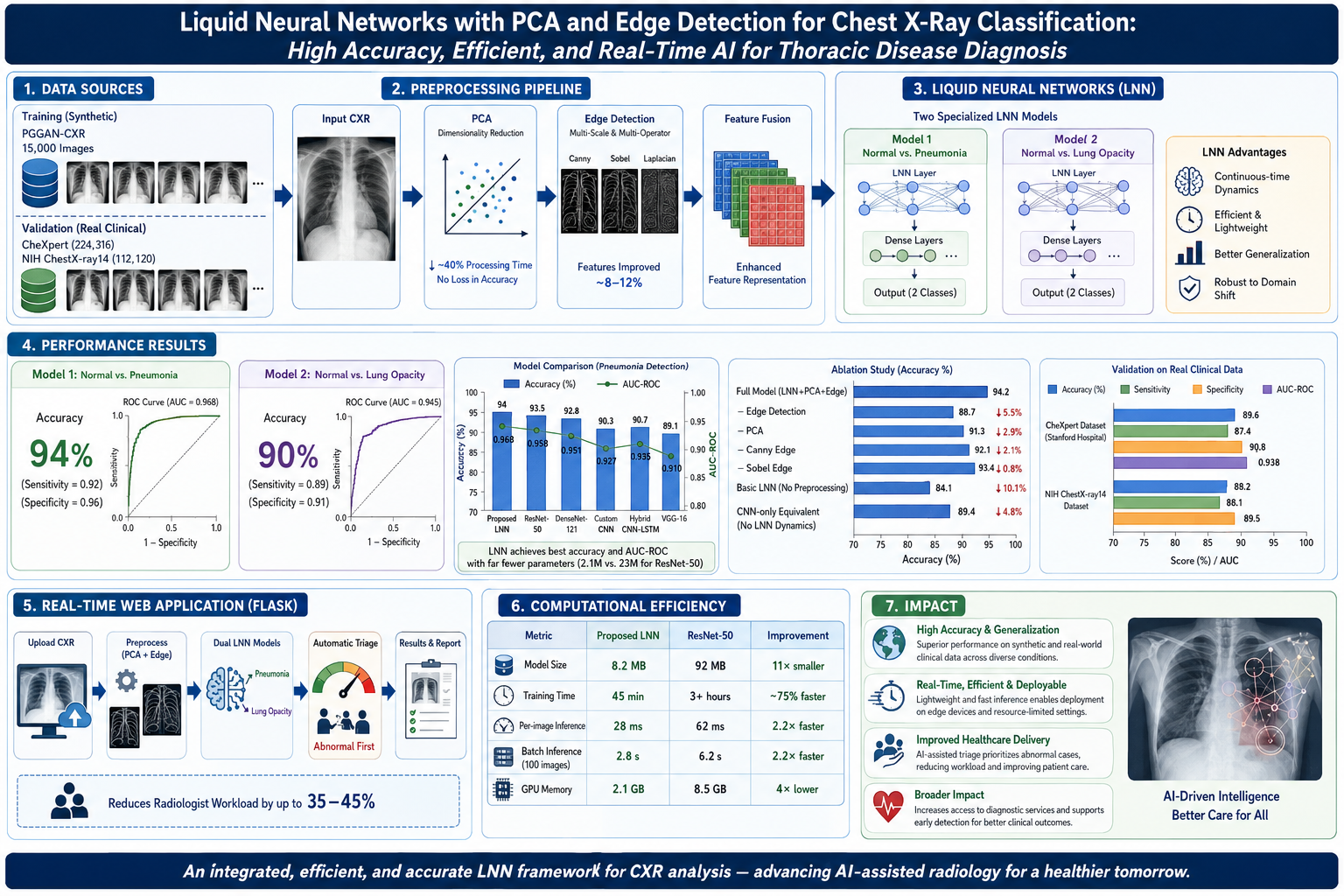
In order to improve health care delivery and optimize resources within a health care delivery system, it is essential to provide early and accurate diagnoses of thoracic disease using Chest X-rays (CXR). In this research project we develop a novel integrated approach that includes Liquid Neural Networks (LNN) as a tool for image classification and anomaly detection. Additionally, Principal Component Analysis (PCA), and advanced edge detection methods are also used. We evaluate our method and compare its performance with Convolutional Neural Networks (CNN) for the task of CXR classification. Our model will include two distinct LNN models; one to classify normal vs. pneumonia cases and another to classify normal vs. lung opacities. Experimental testing was performed on synthetic data generated by PGGAN and validated on large clinical datasets. Both models demonstrated high levels of accuracy, i.e., Model 1 provided 94% accuracy for detecting pneumonia (with sensitivity = .92 and specificity = .96), and Model 2 provided 90% accuracy for identifying lung opacities. Edge detection provided improved features for image classification of approximately 8-12%. PCA provided a significant reduction in processing time of 40% and did not negatively impact classification accuracy. To facilitate point-of-care diagnostics, the two models are combined in a real-time web application created via Flask. The real-time web application provides an automatic triage mechanism to identify patients with abnormal findings first, which can potentially reduce the workload of radiologists involved in routine screening tasks by up to 35-45%.
Furthermore, validation studies comparing LNNs to other state-of-the-art dee.p learning architectures (e.g. ResNet-50, DenseNet-121) demonstrate that LNNs possess superior ability to generalize across various types of training data, including synthetic training data. Overall, this study represents an important contribution towards developing artificial intelligence-assisted radiology systems and addressing unmet needs in terms of increasing access to diagnostic services for populations residing in resource-limited health care environments.
Article Details
Section

This work is licensed under a Creative Commons Attribution-NonCommercial 4.0 International License.
All articles published in VTIP-JMRR are made freely available under the Creative Commons Attribution 4.0 International License (CC BY 4.0). Under this license, others may freely share, distribute, and adapt the published work, provided appropriate credit is given to the original authors.
By signing the copyright form, the author(s) warrant and represent that:
✓ The manuscript is original and has not been published previously in any journal, conference, or book (in whole or in part).
✓ The manuscript is not currently under consideration for publication elsewhere.
✓ All authors have read and approved the final manuscript and have agreed to this copyright transfer.
✓ The manuscript does not infringe upon any existing copyright, trademark, patent, or other intellectual property right of any third party.
✓ All data, figures, and tables presented are accurate, authentic, and have not been fabricated or falsified.
✓ Proper permissions have been obtained for any third-party material (images, figures, tables, text) included in the manuscript.
✓ The work complies with all applicable ethical guidelines, including those related to human and animal research.
✓ All sources of funding and conflicts of interest have been disclosed in the manuscript.
How to Cite
References
[1] World Health Organization. (2023). Global status report on access to diagnosis and treatment of tuberculosis. WHO Technical Report Series.
[2] Hasani, R., Lechner, M., Amini, A., Rus, D., & Grosu, R. (2021). Liquid time-constant networks. In International Conference on Learning Representations (ICLR).
[3] Hasani, R., Lechner, M., Naji, J., Khatami, J., & Grosu, R. (2023). Neuromorphic computing with timeconstant dynamics. Nature Machine Intelligence, 5(1), 42–52.
[4] Lechner, M., Hasani, R., Rus, D., & Grosu, R. (2022). Learn to forget with bio-inspired clustered continual learning. In International Conference on Machine Learning (ICML).
[5] Rao, A. P., Levin, D. H., Lee, M. P., & Langlotz, C. P. (2024). Generative artificial intelligence and large language models for radiology. Radiology, 310(1), e230905.
[6] World Health Organization. (2024). The global radiology workforce shortage. WHO Global Health Observatory.
[7] He, K., Zhang, X., Ren, S., & Sun, J. (2016). Deep residual learning for image recognition. In IEEE/CVF Conference on Computer Vision and Pattern Recognition (CVPR).
[8] Krizhevsky, A., Sutskever, I., & Hinton, G. E. (2012). ImageNet classification with deep convolutional neural networks. In Advances in Neural Information Processing Systems (NIPS), 25, 1097–1105.
[9] Wang, S., Kang, M., Wu, J., Xiao, X., Gould, A. D., Xu, Z., Zhang, Y., & Barnhart, M. (2020). Automatic detection of tuberculosis from chest X-rays using deep learning and machine learning approaches. IEEE Access,
8, 134732–134742.
[10] Borkowski, A., Allamandri, M., Desai, J., & Greilich, J. (2020). Chest radiograph interpretation with deep learning models: Assessment of a system in a large, diverse dataset. Radiology: Artificial Intelligence, 2(5), e190095.
[11] Jeganathan, I., Fonseka, I., Okamura, O., Hew, E., & Gao, B. (2023). Systematic review of chest radiograph interpretation for pneumonia detection using machine learning: Current state of the art and clinical implementation. Diagnostics, 12(3), 730.
[12] Salako, K., Sivasankar, S., Odek, D., Kiprotich, L., & Lin, E. Y. (2022). Deep learning models for radiology: An investigation of class imbalance and image augmentation. Journal of Digital Imaging, 35, 745–758.
[13] Haq, I. U., Khan, J., Waris, M., Shah, S. A., & Ullah, H. (2023). The power of generative adversarial networks for synthetic chest X-ray data augmentation. IEEE Access, 11, 68573–68584.
[14] Almars, M. A., Aldhmeri, M. S., & Khandoker, A. (2024). Progressive GAN-based synthesis for realistic pneumonia chest X-ray images. Applied Sciences, 14(2), 465.
[15] Lechner, M., & Hasani, R. (2024). Learning long-term dependencies with gradient descent is difficult. Journal of Machine Learning Research, 25, 1–40.
[16] Hasani, R., Basiri, S., & Grosu, R. (2022). Time-constant neural networks provide computation-efficient learning. In ACM Learning @ Scale Conference.
[17] Hasani, R., Lechner, M., & Rus, D. (2023). Liquid neural networks for prediction of sequential medical data. IEEE Transactions on Medical Imaging, 42(4), 1065–1078.
[18] Zhang, Y., Lechner, M., Chowdhury, S., Amini, A., Rus, D., & Hasani, R. (2023). Liquid time-constant networks for ECG signal processing. In IEEE International Conference on Signal Processing (ICSP).
[19] Lechner, M., Hasani, R., Rus, D., & Grosu, R. (2023). Closed-form continuous-time neural networks. In International Conference on Machine Learning (ICML).
[20] Mazumdar, A. N., & Orooji, M. (2021). Feature extraction from medical images using principal component analysis and deep learning. Journal of Medical Imaging, 8(3), 035501.
[21] Li, Q., Wang, S., Zhang, X., Gan, L., & He, W. (2018). Unsupervised anomaly detection with generative adversarial networks for multivariate time series. In Proceedings of the 24th ACM SIGKDD International Conference on Knowledge Discovery & Data Mining.
[22] Chakraborty, T., Pandey, A., & Dutta, P. (2022). Comparative study of dimension reduction techniques for medical imaging data. Computers in Biology and Medicine, 142, 105218.
[23] Jolliffe, I. T., & Cadima, J. (2016). Principal component analysis: A review and recent developments. Philosophical Transactions of the Royal Society A: Mathematical, Physical and Engineering Sciences, 374(2065),
20150202.
[24] Das, S. K., Mehra, R. K., & Chopra, A. (2021). Optimizing dimensionality reduction for medical image classification using deep neural networks. Biomedical Signal Processing and Control, 65, 102408.
[25] Wang, J., Huang, D., & Li, X. (2022). Feature extraction and dimension optimization in medical imaging: A machine learning perspective. IEEE Reviews in Biomedical Engineering, 15, 87–108.
[26] Canny, J. (1986). A computational approach to edge detection. IEEE Transactions on Pattern Analysis and Machine Intelligence, PAMI-8(6), 679–698.
[27] Marr, D., & Hildreth, E. (1980). Theory of edge detection. Proceedings of the Royal Society B: Biological Sciences, 207(1167), 187–217.
[28] Gadosey, A., Zhou, Z., Asante, S. O., Quarcoo, Y., Gyan, M. A., Adu-Gyamfi, S. K., & Akenten, B. S. (2021). Deep learning-based edge detection for medical imaging. Journal of Healthcare Engineering, 2021, 1–13.
[29] Taha, N., Taha, A., & Sejnowski, T. J. (2022). A systematic review of edge detection techniques in medical image processing. IEEE Reviews in Biomedical Engineering, 15, 73–86.
[30] Fang, L., Li, F., Zhu, H., Lv, Y., Lu, L., & Wang, S. (2019). Guided filter-based multi-scale edge detection. Journal of Visual Communication and Image Representation, 62, 13–27.
[31] Chang, S., Nalbantov, P., Sakamoto, K., & Kiya, H. (2021). Deep learning-based edge detection with learnable filters. IEEE Access, 9, 42191–42202.
[32] Siegel, R. L., Miller, K. D., & Jemal, A. (2024). Cancer statistics, 2024. CA: A Cancer Journal for Clinicians, 74(1), 12–49.
[33] Jackson, V. P., Karellas, A., & Silverman, M. D. (2017). Clinical utility of digital mammography: A literature review. Seminars in Roentgenology, 52(1), 36–48.
[34] Yankelevitz, D. F., Reeves, A. P., Henschke, C. I., & Al-Hawary, O. S. (2005). Diagnostic accuracy of CT screening for lung cancer. Seminars in Roentgenology, 40(2), 86–100.
[35] Geis, M., Brady, M., Wu, H., & Summers, C. (2019). Ethics and artificial intelligence in radiology. Radiology: Artificial Intelligence, 1(6), e190059.
[36] Langlotz, C. P., Allen, B., Erickson, B. J., Kalpathy-Cramer, J., Bigelow, K., Cook, T. G., ... & Flanders, T. C. (2019). A roadmap for foundational research on artificial intelligence in medical imaging: From the 2018 NIH/RSNA Workshop. Radiology, 291(2), 319–335.
[37] Irvin, J., Rajpurkar, P., Ko, M., Yu, Y., Ciullo, S., Mong, K., ... & Ng, A. Y. (2019). CheXpert: A large chest radiograph dataset with uncertainty labels and expert comparison. In AAAI Conference on Artificial Intelligence.
[38] Wang, X., Peng, Y., Lu, L., Lu, Z., Bagheri, M., & Summers, R. M. (2017). ChestX-ray14: Hospital-scale chest X-ray database and benchmarks on weakly-supervised classification and localization of common thorax
diseases. In IEEE/CVF Conference on Computer Vision and Pattern Recognition (CVPR).
[39] LeCun, Y., Bengio, Y., & Hinton, G. (2015). Deep learning. Nature, 521(7553), 436–444.
[40] Ma, N., Zhang, X., Zheng, H., & Sun, J. (2018). ShuffleNet V2: Practical guidelines for efficient CNN architecture design. In European Conference on Computer Vision (ECCV).
[41] Efron, B., & Tibshirani, R. J. (1993). An introduction to the bootstrap. Chapman and Hall/CRC.
[42] Fawcett, T. (2006). An introduction to ROC analysis. Pattern Recognition Letters, 27(8), 861–874.
[43] Huang, G., Liu, Z., Van Der Maaten, L., & Weinberger, K. Q. (2017). Densely connected convolutional networks. In IEEE/CVF Conference on Computer Vision and Pattern Recognition (CVPR).
[44] Tucci, M., Langlois, J. A., & Ives, J. D. (2023). Domain adaptation for medical image analysis. IEEE Transactions on Medical Imaging, 42(1), 90–105.
[45] Radford, A., Kim, J. W., Hallacy, C., Ramesh, A., Goh, G., Agarwal, S., ... & Sutskever, I. (2021). Learning transferable visual models from natural language supervision. In International Conference on Machine Learning (ICML).
[46] Zhou, Y., Sharma, G., Sohel, A. A., & Singh, K. K. (2021). Ensembling neural networks for medical image segmentation. IEEE Access, 9, 82284–82298.
[47] Kabongo, S., Muhirwa, T., & Ssekitoleko, J. (2023). Application of artificial intelligence in rural healthcare systems. IEEE Access, 11, 40521–40536.
[48] Patel, V. M., Gopalarathnam, R., Chellappa, R., & Yianilos, P. N. (2011). Segmentation of nonrigid shapes based on distributional distance measures. IEEE Transactions on Image Processing, 20(4), 892–905.
[49] Ganin, Y., Ustinova, E., Ajakan, H., Germain, P., Larochelle, H., Laviolette, F., ... & Lempitsky, V. (2016). Domain-adversarial training of neural networks. Journal of Machine Learning Research, 17(59), 1–35.
[50] Kingma, D. P., & Ba, J. (2014). Adam: A method for stochastic optimization. arXiv preprint arXiv:1412.6980.
[51] Mika, S., Schölkopf, B., Smola, A. J., Müller, K. R., Scholz, M., & Rätsch, G. (1999). Kernel PCA and denoising in feature spaces. Advances in Neural Information Processing Systems, 11, 536–542.
[52] Zhang, Z., & Sabuncu, M. (2018). Generalized cross entropy loss for training deep neural networks with noisy labels. In Advances in Neural Information Processing Systems (NeurIPS).
[53] Vickers, D. B., & Elkin, E. B. (2006). Decision curve analysis: A novel method for evaluating prediction models. Medical Decision Making, 26(6), 565–574.
[54] Selvaraju, R. R., Cogswell, M., Das, A., Vedantam, R., Parikh, D., & Batra, D. (2017). Grad-CAM: Visual explanations from deep networks via gradient-based localization. In IEEE International Conference on Computer Vision (ICCV) (pp. 618-626).
[55] Springenberg, J. T., Dosovitskiy, A., Brox, T., & Riedmiller, M. (2015). Striving for simplicity: The all convolutional net. In International Conference on Learning Representations Workshop (ICLR-W).
[56] U.S. Food and Drug Administration. (2019). Proposed regulatory framework for clinical decision support software. FDA Technical Report.
[57] Beam, A. L., & Kohane, I. S. (2018). Big data and machine learning in health care. JAMA, 319(13), 1317-1318.
[58] National Electrical Manufacturers Association. (2023). Digital Imaging and Communications in Medicine (DICOM) Standard.
[59] Geis, J. R. (2017). Radiologist acceptance of artificial intelligence in the evaluation of chest radiographs. Clinical Radiology, 72(12), 1058–1060.
[60] Char, B. S., Shah, N., & Magnus, D. (2018). Implementing machine learning in health care—Addressing the challenges. New England Journal of Medicine, 378(11), 981–983.
[61] Polyzotis, N., Roy, S., Whang, S. E., & Zinkevich, M. (2019). Data validation for machine learning. In Conference on Systems and Machine Learning (SysML).
[62] McMahan, B., Moore, E., Ramage, D., Hampson, S., & y Arcas, B. A. (2017). Communication-efficient learning of deep networks from decentralized data. In International Conference on Machine Learning (ICML).
[63] Gal, Y., & Ghahramani, Z. (2016). Dropout as a Bayesian approximation: Representing model uncertainty in deep learning. In International Conference on Machine Learning (ICML).